One of the most exciting things about archaeology is that you never know what you’ll find until you start digging, and sometimes you don’t know what you find when you find it, and that is exactly the case with this discovery. While digging the shovel test …
Hello, my name is Josie Cowles and I am a junior here at MSU currently working with the MSU Campus Archaeology Program (CAP) to excavate the old MSU observatory. The foundation was found by the CAP crew in May of 2023, and has been the …
Hi, my name is Katie Simonson and I am one of the students taking part in the 2024 field school, where we are working on the site of the original observatory here on MSU’s campus. Part of the foundations were found earlier in May of 2023 by CAP, so we are expanding on the work already done. Our field school started on the 28th of May this year, and we went over the history of the site, safety, and got to know our team. We continued this on the next morning, but after that we began the pedestrian survey of the site.
A pedestrian survey is when we walk along the surface of the site to find artifacts on the surface. Since the site is in an area where many people occupy, that means the surface has been very disturbed since the time of the site and it is unlikely we would find any historic artifacts, but it is still important to look. We all walked along a transect, a straight line across the site, that was spaced out a meter apart, and flagged any artifacts we saw. Since the majority of us have never done a pedestrian survey before, we decided to flag all man made objects as practice. After we flagged the objects, we would map their position, take a picture of it, and describe the object using a program called KoboToolbox. If we found anything possibly historic then we put in an artifact bag, otherwise we would dispose of it.
As we predicted, we didn’t find many artifacts that could be historic. We found lots of modern trash including plastic wrappers, cardboard, paper cups, and cans. We also found a rodent bone. Some of the things we found were not historical but still interesting, so were kept by some of us for our own use. For example, I kept a broken cd and a beaded chain to make into a suncatcher. We did find some objects that could possibly be historic, such as pieces from glass vessels, metal bottle caps, and some pieces which were most likely from a dining hall plate. These were bagged for further analysis later.
The pedestrian survey might not have given us much insight into the history of the site, but it did allow us to become familiar with the area and the process of surface survey. On other sites which are less disturbed by modern human activity, it would be more likely to find more historic objects. It is important to know how much a site has been disturbed, because that can affect how we need to study the site.
I hope you enjoyed learning about the pedestrian survey of the 2024 observatory site, and if you are interested in learning more there are plenty of resources on this website about the history of the site and our work here.
A photo of the suncatcher I made using a broken cd and a green beaded chain found during the pedestrian survey.
The importance of archaeological excavations revolve around the drive to uncover forgotten, and missing pieces of history; my time with Michigan State’s Campus Archaeology Program aided in doing just that. I worked alongside Dr. Stacey Camp and 12 other CAP crew members to dig up …
This academic year has allowed me to explore several digital methods I had little to no knowledge about. This is partially due to my teaching position at MSU in the Lab for the Education and Advancement in Digital Research (LEADR). While in this position, I …
This past summer has been one of the busiest, if not the busiest, summers of my time as director of the MSU Campus Archaeology Program. While we did not have a field school as we generally run them every other year, a remarkable discovery was made in May of 2023. As construction workers were installing hammock poles near Wills House, they hit a concrete platform. Thankfully, we work closely with Infrastructure, Planning, and Facilities (IPF), who oversee all construction projects on campus.
We were then contacted by Landscape Architect Yun Cao, who told us about the hammock poles and asked if we had additional information about the history of this particular area of campus. Ben Akey, our current Campus Archaeologist, examined our historic maps, historic aerial photographs, and GIS database to reconstruct the landscape’s alterations and changes over time. Ben discovered that MSU’s first observatory dating to circa 1880/1881 was close by, and then investigated the area by conducting shovel tests, or small holes in the ground designed to locate artifacts and architectural features swiftly.
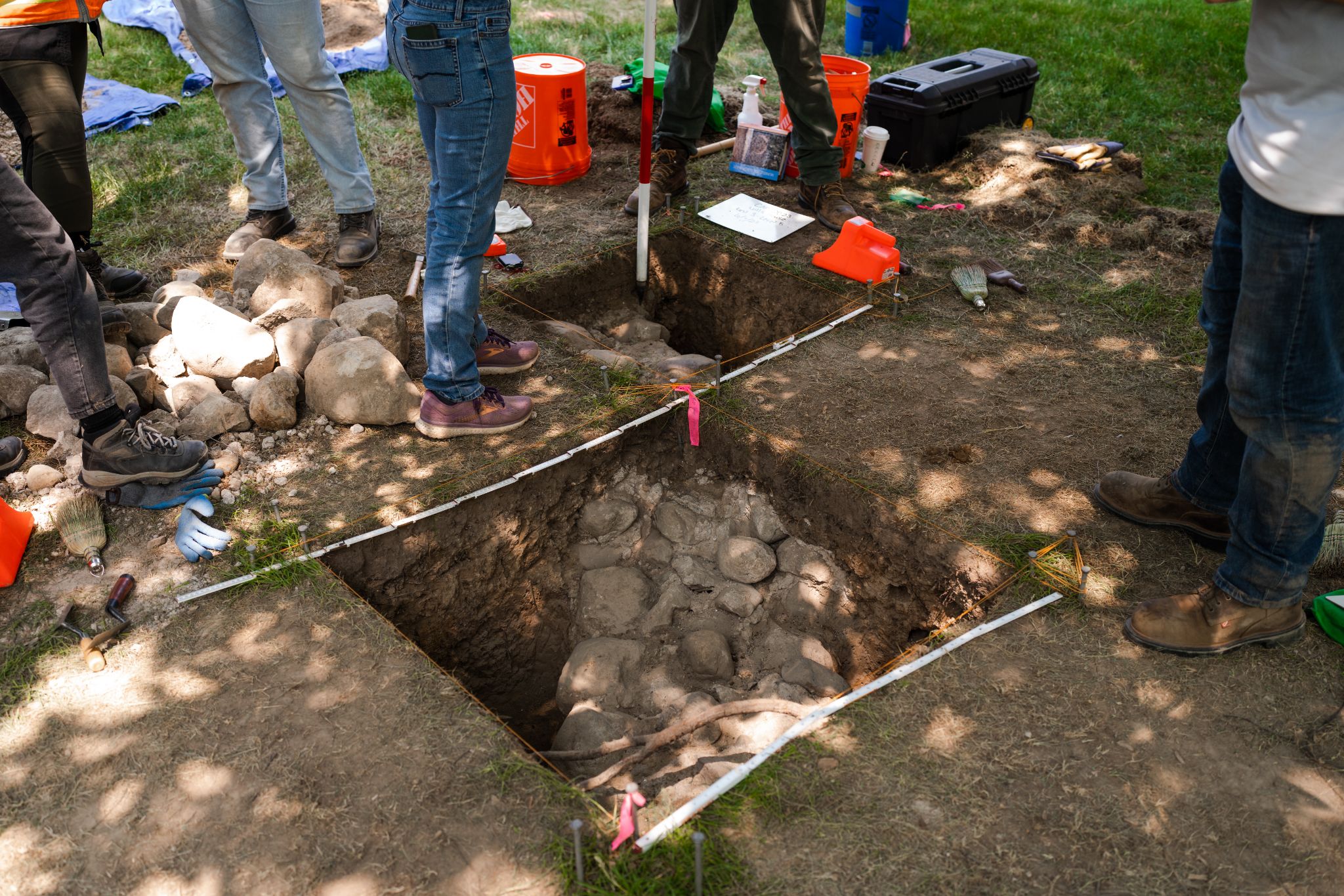
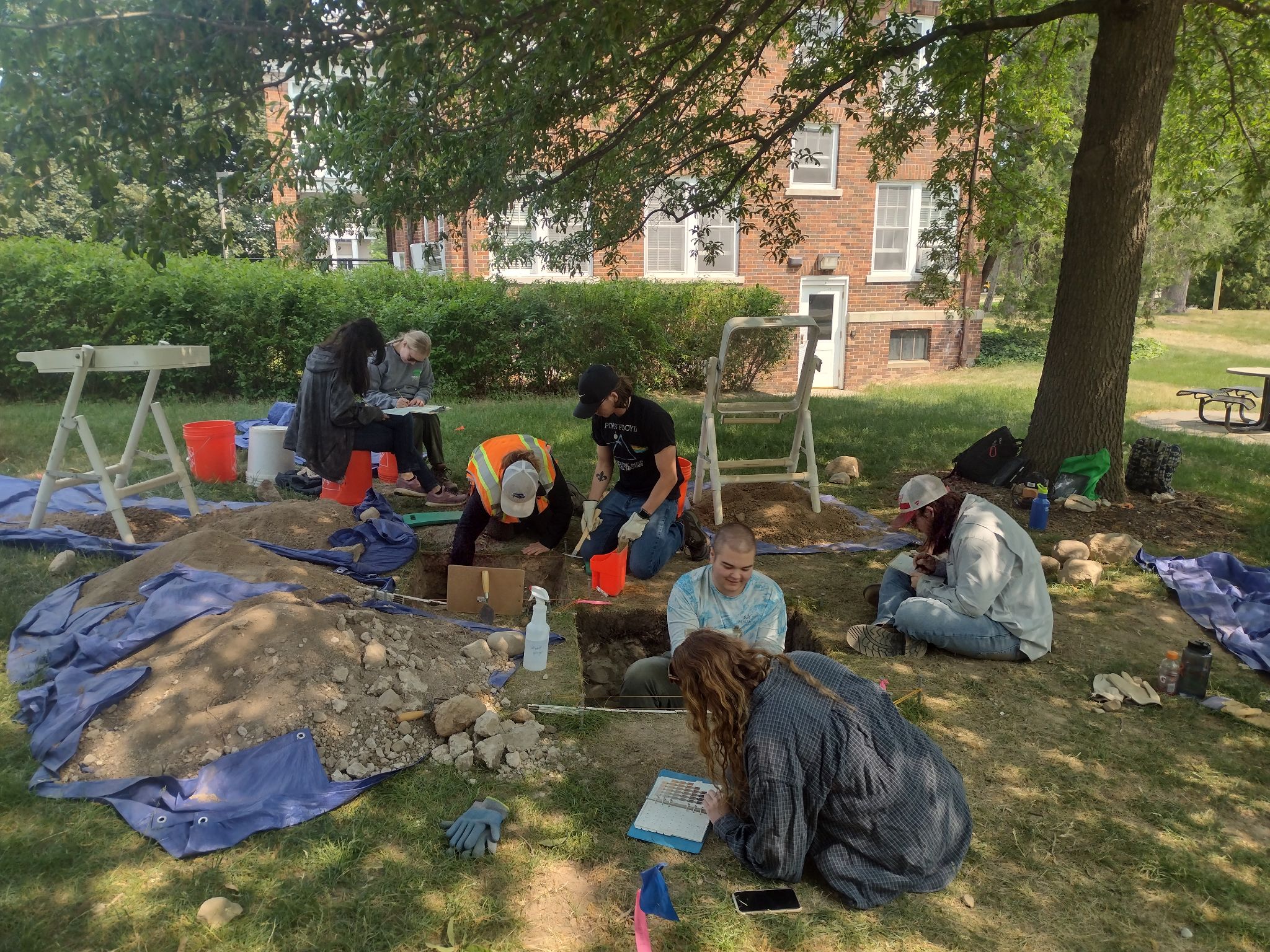
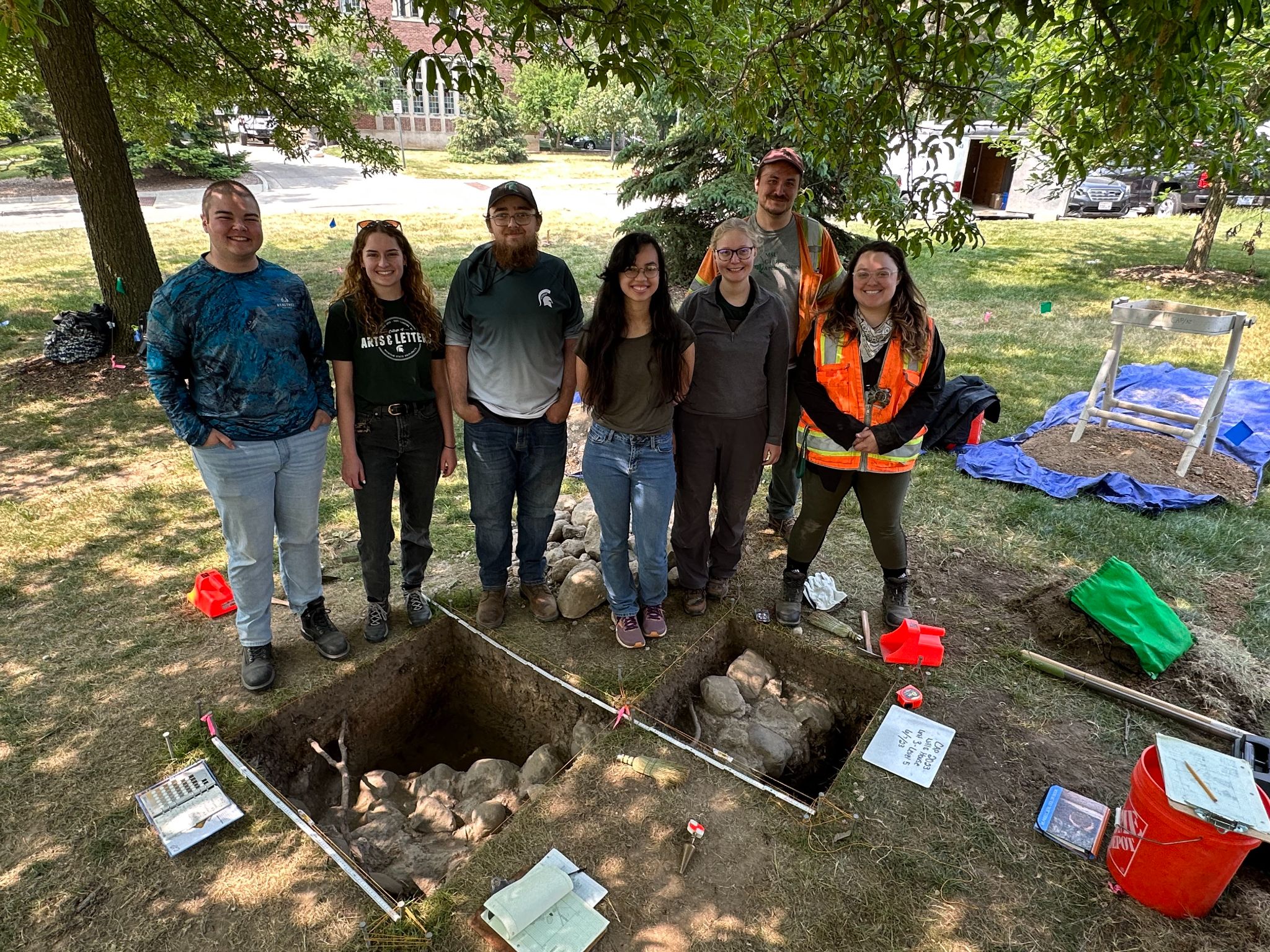
The shovel tests revealed a number of architectural-related artifacts, such as nails, glass, stones, and brick fragments. While this material culture is commonly found across our campus due to substantial construction over time, Ben had the foresight to open up 1×1 meter excavation units to further explore the structural materials. As a result, portions of the original foundation were uncovered, leading to an explosion of interest from the media and our campus community! Below you can see the two 1×1 meter units that exposed the observatory’s foundation and our team of graduate and undergraduate students excavating it.
Since we had several other critical construction projects taking place in the summer, we decided to close up the units and move to our other sites. We made this decision because we knew the site was not slated for construction and would not be destroyed over the next year. We knew that this would be a great place to study as part of an undergraduate field school, too, so we decided to carefully backfill the excavation units with dirt, using materials to protect the foundation in the process.
This summer (2024) we will be returning to the site, and undergraduate and graduate students will have the opportunity to excavate the remaining portions of the foundations. We will also have the chance to try to locate other features from this time period; perhaps we will find an outhouse that was used by the students who studied in the observatory, or Professor Rolla Carpenter himself! More information about the field school can be found on our website here.
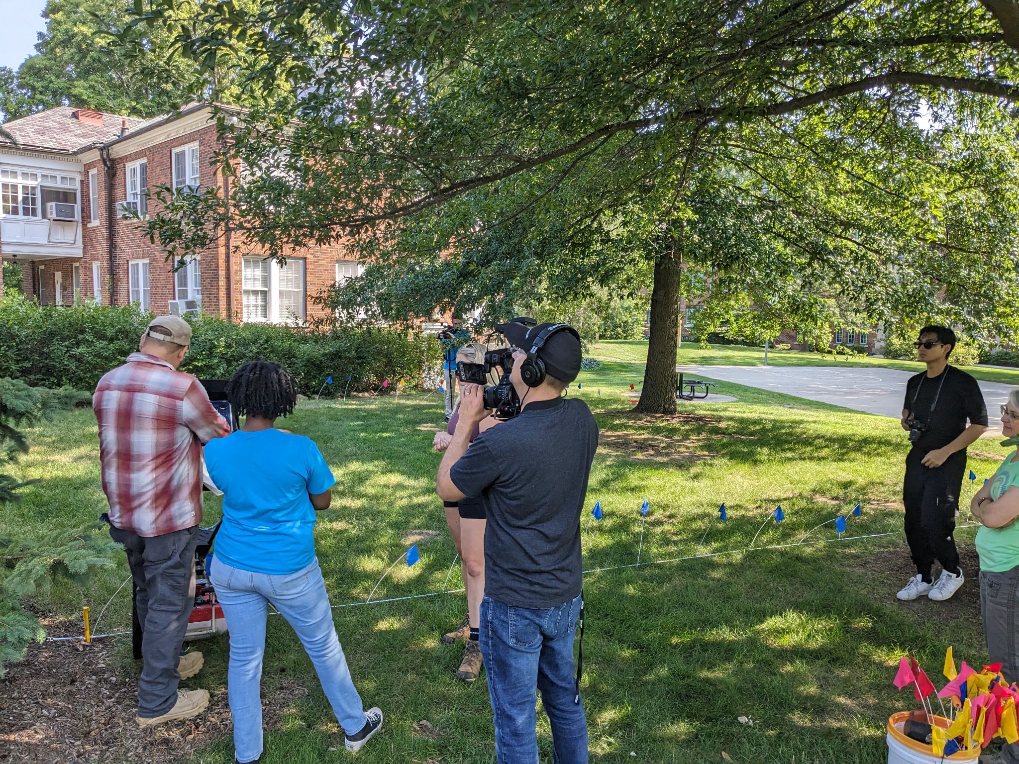
One of the questions I wanted to answer before the summer ended was if the entire circular foundation was intact. Dr. Duane Quates (an alumni of our program) and Dr. Chris Valvano (also an alumni!) kindly visited the site and conducted a ground penetrating radar survey (GPR) of it. They just so happened to be there during a day when PBS/WKAR was filming a show on archaeology and STEM with a teenage actress, who was able to get to try out GPR!
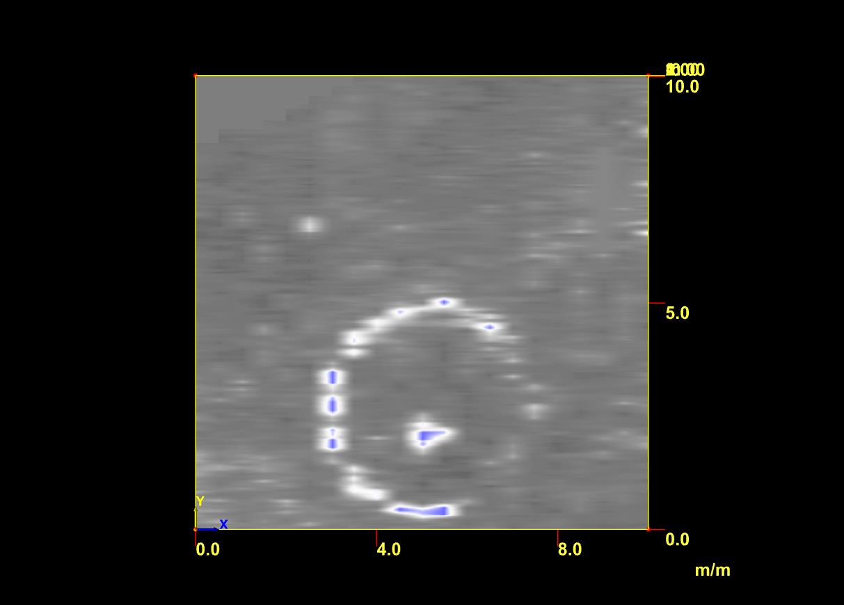
I learned a bit about what it was like to be on television, and we also learned more about what was literally hidden beneath our feet. While the GPR data is preliminary, it appears as though most of the observatory’s foundation is intact (see image of the GPR data below). We will find out more when we return to excavating in late May of 2024.
As you can tell from the GPR data, the foundation was circular, about 16 feet across. Archival records also note that the observatory was this size. There is a unique feature in the middle of the observatory’s foundation. It appears as though there may be a platform for the telescope originally housed in the observatory. This is a common for observatories, as the foundation is intentionally separate from that which stabilizes the telescope.
Since we all recognized what a remarkable discovery this was, we immediately shared our finding with communications and public relations staff at MSU. Alex Tekip, Sydney Hawkins, and Nick Schrader came out to the site immediately, taking photographs, videos, and interviewing our staff on site. I knew this was an important story, but I did not realize that this would become an international story nearly overnight! In July, we were fielding media requests daily, which was a learning experience for myself and my students. We received advice and help from Alex, which was greatly appreciated. Thanks to MSU PR’s hard work and detailed write-up of our discovery, we were featured in national and international media outlets, including People Magazine, the New York Times, the Washington Post, NPR, USA Today, the Guardian, Popular Science, and Space. Interest in the site hasn’t died down, as Smithsonian Magazine just named us one of the most 117 interesting and exciting discoveries of 2023!
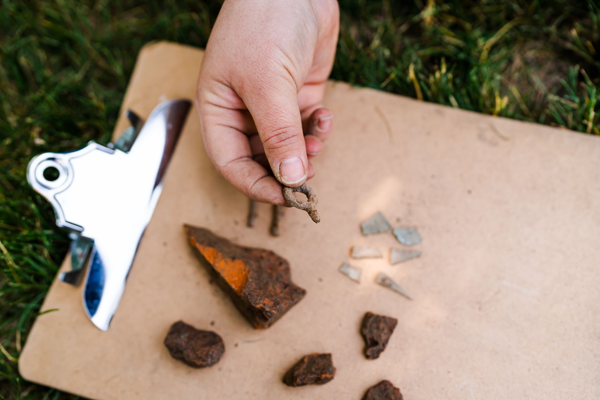
To help facilitate additional research on the observatory before we hold our field school this summer, I was able to obtain a Provost’s Undergraduate Research Initiative (PURI) grant for one of my undergraduate students, Hannah. This will allow us to clean and catalog the artifacts found during last summer (see the image of some of them below), and locate additional archival documents and images related to the observatory. Believe it or not, with the help of MSU University Archives & Historical Collections Hannah has already found a previously undiscovered image of the observatory, revealing another mystery. This photo below illustrates what looks to be a small bench or structure above ground. We have talked to a number of folks about it, including MSU Professor Emeritus (of Astronomy and Astrophysics) Dr. Horace Smith, who wrote a book on the history of astronomy at MSU. There are a lot of ideas as to what this feature is – we hope to solve this mystery in the summer!
Image courtesy of Jenny Rankin and Hannah Magnus. Found by Hannah Magnus at the MSU University Archives & Historical Collections, UA 10.3.65 Charles Philip Close papers, Scrapbook #62 (dates from 1891-1895). Photo identifier A010539.
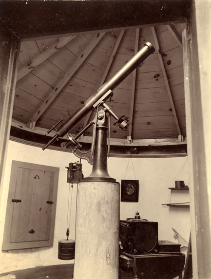
There is still a lot of work to be done at the observatory site, but we are writing up a long summary of it that will hopefully be published in the near future. The observatory was built with the assistance of student labor, a common practice in MSU’s earliest days. Professor Rolla Carpenter, pictured below, was also was able to obtain funding for the telescope inside the observatory, which now sits at MSU’s Abrams Planetarium in an exhibit. It is incredible to think that the original telescope inside the observatory is still alive and well in our planetarium!
The only known photograph of the interior of MSU’s first observatory, with telescope in the center. This telescope is currently on exhibit at MSU’s Abrams Planetarium. The podium in the center might be the circular feature identified via the GPR survey. Photo courtesy of MSU University Archives & Historical Collections. Resource identifier A000122.jpg. Photo circa 1909.
Dr. Shannon Schmoll, Director of the Abrams Planetarium, and I are working to coordinate a more robust exhibit at the Abrams Planetarium that will include artifacts and new photographs. It is a work in progress, but we hope to share more about it later this year. Shannon, her intern, Caitlyn Tanner, and I have had a couple of adventures this past year to learn more about the observatory and the people who used it. We visited Detroit Observatory in the fall, which dates to 1854 (a bit earlier than MSU’s observatory, and a lot more grand in size!), and then some of MSU Museum‘s enormous textiles collection in late fall. The textiles collection gave us a sense of what students and professors wore on campus in the 1880s and 1890s. We were curious if we might be able to see clothing similar to that which students wore in the MSU observatory photograph below.
Photo courtesy of MSU University Archives & Historical Collections, Collection Number UA 10.3.265. Photo circa 1888. Professor Rolla Carpenter, who helped obtain funding for the observatory and taught classes in astronomy at MSU, is pictured in the center of the photograph, tipping his hat.
We have so many questions about the observatory. Hannah, our PURI grant recipient, is spending hours in MSU’s University Archives & Historical Collections looking for previously undiscovered photos of the observatory (which she already discovered on day 1 of being in the archives!). She is hoping to identify some of the people in the above photograph. Who is the sole woman in the photograph? In the late 1800s, it was unusual for a woman to have access to a university education. We presume she may have been the daughter of a faculty member on campus. And what about the men? Who are they? There weren’t many students at MSU during this time period, so hopefully we can identify each and every individual photographed – if we are lucky, maybe we will even located their descendants. Maybe some of them are walking on this campus as I write 🙂
We are very thankful to the crew of MSU students who were a part of this incredible discovery. These students include (from left to right of the image below: Levi (undergraduate student), Morgan (undergraduate student), Mac (undergraduate student), Kelly (undergraduate student), Holly (Ph.D. student), Ben (Campus Archaeology, Ph.D. student), and Tori (Ph.D. student)). If it wasn’t for student labor, the observatory wouldn’t have been built. And if it wasn’t for student labor, we wouldn’t have discovered the observatory 143 years later!
by Juan Carlos Rico Noguera Michigan State University (MSU) CAP “is a program that works to mitigate and protect the archaeological resources on Michigan State University’s beautiful and historic campus.”[1] CAP is also an initiative that contributes to the public understanding of MSU’s history, enabling …
Holly Long I love tea; I drink it every single day. It is warm, hydrating, and is known for healing properties. But the tea leaves most drink today are imported and are not indigenous to North America and are rarely grown here. Tea leaves, not …
Construction along Service Road in 2020 found a mid-20th-century midden. The artifacts found were associated with the history of temporary post-World War II student housing on Michigan State’s campus. After the Servicemen’s Readjustment Act of 1944, or the GI Bill, became law, college enrollment increased in the United States (Dressel 1987). This required more housing on campus, especially for married students, many of whom had families (Offices of Board of Trustees and President 1944).
During the summer of 2021, I worked on the Campus Archaeology summer crew. On rainy days, we worked in the lab, cleaning and cataloging materials from Service Road. While cataloging, we noticed a lot of underglaze, decal-decorated institutional-ware ceramics with similar patterns. By the end of the summer, we identified four distinct decorative patterns, which we named MSU Green Band, Esquire, Mobile, and Cross Stitch. These would have been used in dining halls, and their decorative styles allowed us to learn more about the places on campus where these ceramics would have been used. Additionally, we were able to find distinct dates for the ceramics based on the maker’s marks and date codes, when present. Date codes allow for a level of granularity in our analyses that is rare with other kinds of artifacts, providing a means to trace shifting patterns of institutional requisition.
Ceramic plate with the Shenango China Company maker’s mark. The date code is indicated by the red arrow.
MSU Green Band
Plate with MSU Green Band design with the Michigan State University logo.Bowl with MSU Green Band design with the Michigan State College logo.Creamer with MSU Green Band design.
The MSU Green Band design is named for the single green line just below the rim. Additionally, on some vessels the institution’s seal is located just below the green band. This pattern was used on dining hall dishes for much of the 20th century. Based on our preliminary analyses, it appears this design may have been gradually replaced by the Esquire pattern.
Two versions of the MSU Green Band design are pictured. As Michigan State shifted from College to University in 1955; the MSU Green Band design shifted as well, which can be seen reflected in the two distinct seals pictured in the examples provided above. MSU seems to have ordered this design from multiple companies, and the examples in the Service Road collection were produced by either the Shenango China Company or the Mayer China Company. MSU Green Brand was the most enduring ceramic style in the Service Road collection, with maker’s marks indicating a date range of 1950 to 1963. The MSU Green Band design is the most represented of the four ceramic types, comprising the majority of ceramics recovered from the site.
Esquire
Mug with a broken handle, the Esquire design runs below the rim.
A second decorative pattern identified in the Service Road collection has been named ‘MSU Esquire’. The Esquire pattern takes its name from a similar Shenango China design called “Esquire.” Relative to the original Shenango design, the vessels recovered from the Service Road landfill had rectangular spiral designs rather than squares, and the laurels extend along longer stretches of the design (Replacements 2021). It seems likely that MSU commissioned a distinct version of the Esquire pattern for the university, though we have not been able to locate records to corroborate this. Our preliminary analyses suggest this design may have gradually replaced the MSU Green Band design.
Mobile
Cup with the Mobile design running from rim to the center of the body.
The Mobile design consists of a fading grey band along the rim and a singular black and grey baby “mobile” motif that then extends from the rim to the larger undecorated portion of the vessel. The Shenango China Company created these ceramics, and the dates for this vessel range from 1951 to 1961. This pattern was created specifically for MSU’s new Kellogg Center for Continuing Education (Pratt 2003:116).
Cross Stitch
Cup with the Cross-Stitch design running below the rim.
The last design found was Cross Stitch (Arthus 1955). The pattern consists of squares arranged in a floral motif, resembling traditional cross-stitching patterns. The stems and leaves are green, with blue and red alternating flowers; these designs run below the rim. The Cross Stitch design had Shenango and Mayer China maker’s marks. Compared to other designs discussed here, we recovered substantially fewer examples of the cross-stitch pattern. The few finely-dateable examples of this pattern in the Service Road collection were produced between 1958 and 1959, though we know from the archival photo below that use of this pattern at the university extends back to at least 1948.
Women being served on Cross-Stitch design ceramics, which are circled in red. Dated January 15th, 1948. Photo courtesy: MSU Archives and Historical Collections.
This archival photograph of dining service at Landon Hall features this pattern, suggesting that it was used in women’s dining halls alongside other patterns like the MSU Green Band (also pictured). Given this photographic evidence and gendered imagery incorporated into the design, this pattern may have been exclusive to women’s dining halls (Michigan State University 1960; UAHC 2021).
Final thoughts
The ceramics found in the Service Road midden were used in several distinct areas of campus, ranging from dining halls to the Kellogg Center. The abundance of complete and near-complete dishes in the Service Road collection allowed us to begin serializing ceramics used on campus in the mid-twentieth century. Being able to identify different ceramic designs utilized across MSU’s campus supports future CAP research efforts, as we now have a better sense of when and where on campus they were utilized.
References
Arthus, Gerard (1930) Mayer China: Illustrated Book of Decorations, No. 10. Mayer China Company, Beaver Falls, PA.
Dressel, Paul (1987). College to University: The Hannah Years at Michigan State, 1935-1969. Michigan State University Press, East Lansing, MI.
Michigan State University (1960) “The Helot: Student Handbook”, Michigan State University Publications, East Lansing, Michigan. Available online, https://onthebanks.msu.edu/Object/162-565-2184/student-handbook-1960/, accessed December 23, 2021.
Offices of Board of Trustees and President (1944) Meeting Minutes, December 21, 1944. UA 1. University Archives and Historical Collections, East Lansing, Michigan.
Pratt, Michael E. 2003. Mid-Century Modern Dinnerware: A Pictorial Guide: Red Wing to Winfield. Atglen, PA: Schiffer Pub.
This last summer, I had the amazing opportunity to be a part of CAP Crew, the group of MSU Archaeology (or archaeology-curious) students that conduct the compliance archaeology during the summer. Although, there is significantly more paperwork and lab work than there is fieldwork – …


